 English
 Français
 Deutsch
 euskara
 Русский язык
 Italiano
 Português
 Nederlands
 Polski
 Greek
Greek Lietuva
 Türkçe
 日本語
日本語 한어
한어 中文
中文 தாமில்
 فارسی
فارسی हिंदी
हिंदी Tiếng Việt
 ภาษาไทย
 Pilipino
 Indonesia
 தாமில்
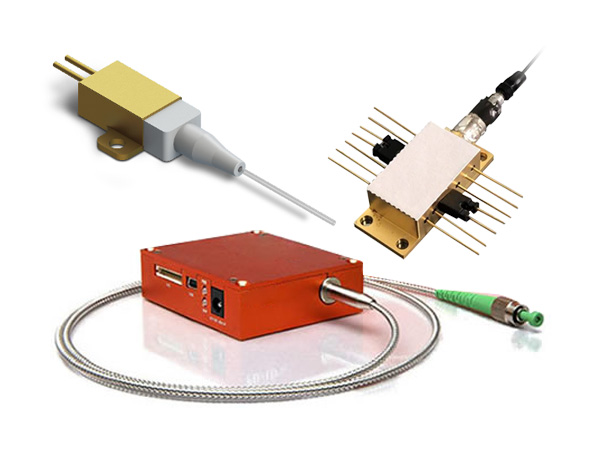

Lasers for Raman Spectroscopy and Microscopy
The energy for this wavelength change comes from a change in the energy state of a molecular bond or bonds. This is distinct from an interaction where the photon is absorbed by an atom and then re-emitted at a different wavelength, which is the domain of fluorescence spectroscopy. The wavelength shift in the Raman scattered light corresponds directly to the current energy states of the molecular bonds in the sample so, as these are influenced not just by the atoms involved in those bonds but also by the total crystal structure and the strain the system is under, it is possible to interpret useful information from the Raman spectrum that can be difficult to obtain by other means.
First demonstrated by C.V. Raman in 1928, this weak Raman effect has been historically difficult to discern. However, significant advances in laser sources, detectors, and optical techniques mean that Raman Spectroscopy and Microscopy are now widely used over a disparate range of applications; including biology, pharmaceuticals, semiconductors, forensics, security, and the analysis of art and museum artifacts.There are several considerations when selecting an appropriate laser source for Raman Spectroscopy:
1. Wavelength
The strength of the Raman signal is directly dependent on the wavelength of the laser source, where lower wavelengths will produce stronger Raman signals, as well as allowing for higher spatial resolution. It is important, however, to balance this observation with the occurrence of background fluorescence, prevalent in many materials throughout the UV-visible spectrum, and the possibility of sample damage at high energy. These effects most often cause a compromise in the wavelength of the source used, where longer wavelengths, such as 532 nm, 785 nm, and 1064 nm, in combination with highly sensitive detectors, allow for the widest range of samples to be measured.
2. Spectral Linewidth and Purity
The spectral linewidth of the laser source should also be considered, as it will limit the possible resolution of the Raman measurement and so the minimum energy change that can be determined. It is important for the laser selected to have a linewidth below the overall resolution of the Raman spectrometer, on the order of picometers, but for high-resolution spectroscopy this is critical, requiring linewidths below 1 MHz. High spectral purity will also increase the signal to noise ratio from the measurement.
3. Beam Quality
The beam quality is related to the possible spatial resolution. Here, single transverse mode beams (TEM00) are vital for confocal Raman Spectroscopy in particular, allowing for high spatial control in all three axes, improving spatial resolution, and decreasing background effects.
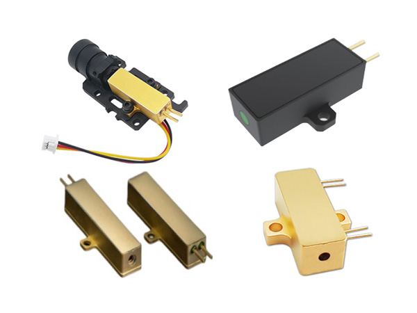
We design and manufacture STU series single frequency laser sources with unrivalled wavelength stability, narrow linewidths and long coherence lengths over a range of wavelengths within a small footprint. We currently offer single-frequency lasers in the red and green areas of the visible spectrum with our Solo 640 Series and Duetto 532 Series respectively; to complement our Solo 1064 Series, for the elimination of background fluorescence and the analyses of biological samples.
For more information on STU series single frequency lasers, please click here.